
Brain Fog After COVID-19
Punita Lalchand (edited by Anindro Bhattacharya)
Posted on 13 Mar, 2023
Introduction
A whopping 20-60% of patients experience long COVID after having a SARS-CoV-2 infection (Novak et al., 2021). SARS-CoV-2 is the virus that causes COVID-19, an acute disease, meaning it lasts for a short period (days to weeks) (Novak et al., 2021; Brutto et al., 2021; CDC, 2022). However, there are symptoms that can occur beyond this period of time, known as the post-acute sequelae of COVID-19, or long COVID (Novak et al., 2021; Brutto et al., 2021). This can manifest differently for everyone, including fatigue, dyspnea and brain fog (Novak et al., 2021). Here, the link between COVID-19 and brain fog, specifically on its epidemiology, risk factors, mechanism, and how it can be prevented will be a central focus. Brain fog occurs when patients who have COVID-19 experience issues with memory, multitasking and sleeping disorders (Sklinda et al, 2021). This is an important concept as brain fog is an example of psychiatric and neurological problems, which can manifest in approximately one-third of those who survive the COVID-19 disease (Sklinda et al., 2021).
Overview of Brain Fog
COVID-19 symptoms can last anywhere from four to twelve weeks, while long COVID can occur after twelve weeks (Callan et al., 2022). Symptoms of brain fog can look different for everyone. Some common ones are issues with executive function, attention, memory, and language (Callan et al., 2022). Lowered maximum grip strength and gait speeds are some examples of these issues (Asadi-pooya et al., 2022). In a cohort study of 2696 people, it was estimated that of those aged 18-55 who contracted COVID-19 and were hospitalized, 7.2% developed brain fog (Asadi-Pooya et al., 2022). Among long COVID patients with neurological symptoms, brain fog is present in 81% of cases (Mathern et al., 2022). It was noted that brain fog was more likely to be present if a patient was female, had respiratory problems the infection started, and intensive care unit (ICU) admission was necessary (Asadi-Pooya et al., 2022). It can occur even in patients with mild symptoms of COVID-19 (Brutto et al., 2021). Cognitive decline was shown to improve over time, twelve months post-COVID-19 infection (Brutto et al., 2021). However, there are currently limited studies on the prognosis of cognitive decline, so it cannot be determined with certainty (Brutto et al., 2021).
How COVID-19 Works
COVID-19 is transmitted through droplets and aerosols, which are tiny particles in the air (Parasher, 2021; CDC, 2010). It has four different proteins that make up its structure, which is its spike (S), membrane (M), envelope (E) and nucleocapsid (N) proteins (Praasher, 2021). These are visualized in Figure 1. The S protein has two subunits, S1 and S2 (Parasher, 2021). The S1 subunit plays a role in attaching to the receptor of the host cell, while the S2 subunit allows for the fusion of the viral and host membranes (Parasher, 2021). The S protein binds to the Angiotensin-converting enzyme-2 (ACE-2) receptor, which is an enzyme that breaks down angiotensin and regulates various cellular functions (Parasher, 2021; National Cancer Institute, n.d.)). ACE-2 receptors can be found in many organs such as the lung, brain, heart, liver and kidneys (Salamanna et al, 2020). These are therefore key areas of infection (Salamanna et al., 2020). ACE-2 is expressed in the pulmonary epithelial cells, and so the virus binds there (Parasher, 2021). The virus fuses with the pulmonary alveolar epithelial cell membranes, which are membranes of alveoli (air sacs) present in the lung, and releases its contents (Parasher, 2021; National Cancer Institute, n.d.). It is then able to replicate using the host cell’s machinery, and the new viral particles leave the cell through exocytosis (Parasher, 2021). Once it leaves, it can go on to infect other cells in other parts of the body (Parasher, 2021). This process can be seen in Figure 2. The immune system then releases different cytokines (proteins that control the growth and activity of other immune and blood cells) and inflammatory markers (proteins released during inflammation that can act as markers for inflammation) to fight the virus (Parasher, 2021; American Cancer Society, 2023; Pearson, 2022). However, they can also lead to inflammation and lung injury, especially with persistent inflammation (Parasher, 2021).
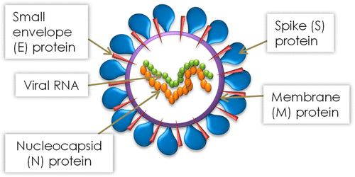
Figure 1: Structural proteins of SARs-CoV-2. Adapted from Gil et al. (2020).
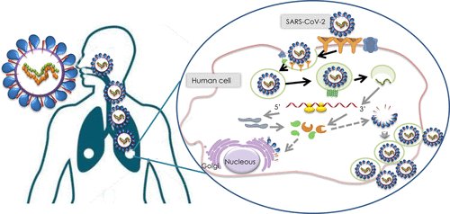
Figure 2: How SARS-CoV-2 infects the body. Adapted from Gil et al. (2020).
How Brain Fog Occurs After COVID-19
The mechanism for how COVID-19 causes neurological injury has not been identified (Mathern et al., 2022). However, there are a few potential pathways that give rise to this phenomenon. One such pathway is through neurons (cells that transmit information throughout the brain and nervous system) and astrocytes (cells that are important for development and more) (Mathern et al., 2022; National Institute of Neurological Disorders and Strole, 2022; Sofroniew & Vintners, 2010). The ACE-2 receptor is expressed on astrocytes and neurons, which is what the COVID-19 spike protein binds to in order to infect a host (Mathern et al., 2022). Neurons are more susceptible to this due to the high resistance to SARS-CoV-2 shown by astrocytes (Mathern et al., 2022). Another potential pathway is through the body’s inflammatory cascade, which is a series of immune mechanisms that repairs tissues and is activated by detecting the virus (Mathern et al., 2022; Megha et al., 2021). This leads to vasculitis (inflamed blood vessels) in the central nervous system, which is a branch of the nervous system made up of the brain and spinal cord (Mathern et al., 2022; Mayo Clinic, 2022; Hirsch, 2022). Another explanation is that the virus causes an autoimmune response, which attacks brain cells instead of the virus (Mathern et al., 2022). These pathways all lead to neurological injury, which can significantly affect someone, such as brain fog, headache and numbness/tingling (Mathern et al., 2022).
SARS-CoV-2 also increases the C-C motif chemokine ligand 11 (CCL11), which is responsible for recruiting eosinophils, a type of white blood cell, to inflamed sites (Li et al., 2021; Foti & Locati, 2017; Cincinnati Children’s Hospital, 2022). This increases the white matter microglial reactivity, which activates microglia, a type of cell involved in the immune response, development and more (Li et al., 2021; Bachiller et al., 2018). This decreases hippocampal neurogenesis, which is the production of new neurons in the adult brain, a process important for memory and learning (Li et al., 2021; Costa et al., 2015). This subsequently decreases the number of neurons (Li et al., 2021). Increased CCL11 also decreases the number of oligodendrocyte precursors, which regenerate oligodendrocytes and myelin (Li et al., 2021; Kirby et al., 2019). As a result, the number of oligodendrocytes, a type of cell responsible for myelin formation (on the axon of neurons), is reduced (Li et al., 2021; Kipp, 2020). This reduces the number of myelinated axons, which is necessary for effective communication between neurons (Li et al., 2021; Papuc & Rejdak, 2018). Decreased myelinated axons have been linked to cognitive impairment, and can potentially explain brain fog in COVID-19 patients (Papuc & Rejdak, 2018). This also decreases the number of neurons in the brain, which can lead to cognitive impairment via brain atrophy (Li et al., 2021; Avila et al., 2017). This cascade is visualized in Figure 3.
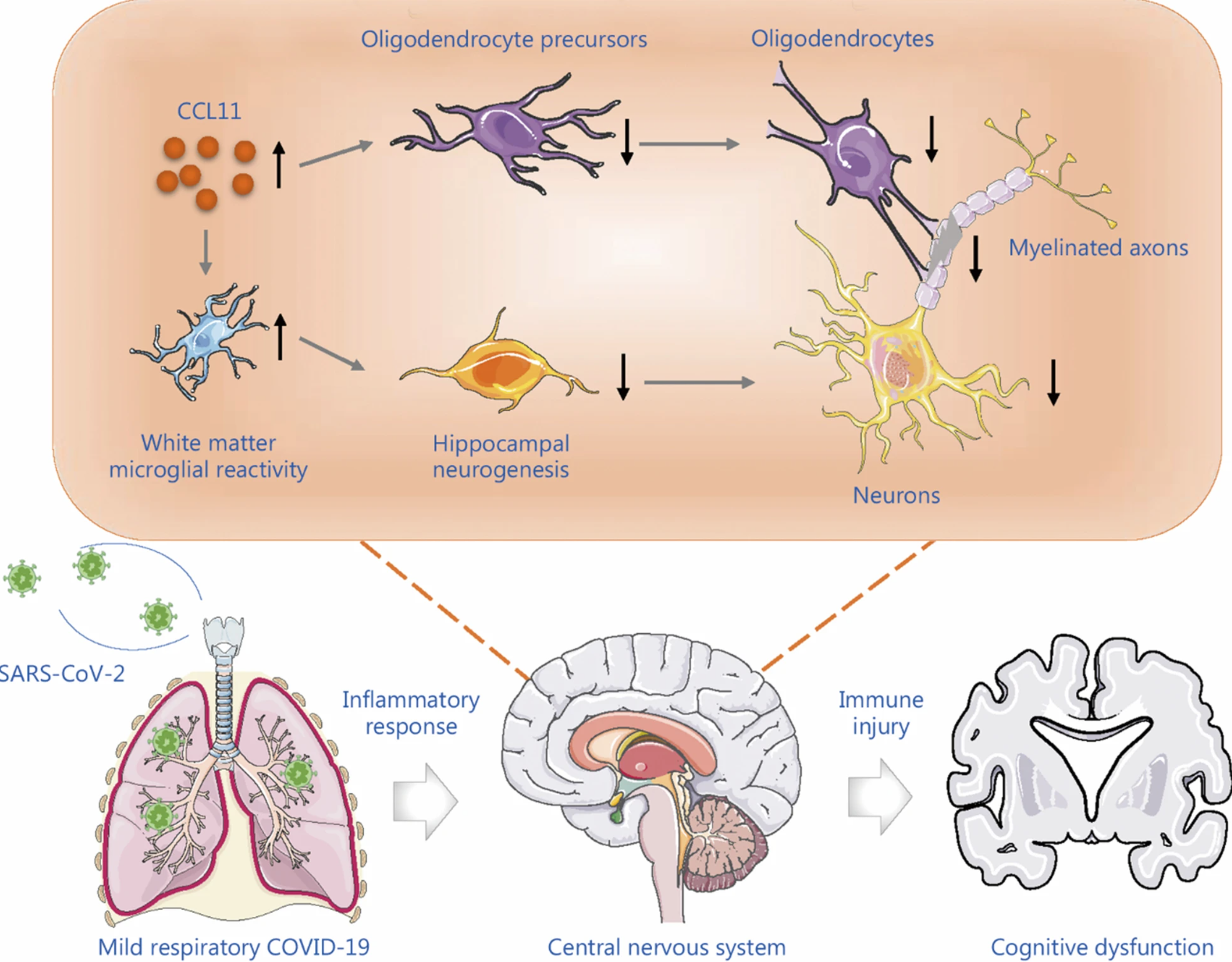
Figure 3: How SARS-CoV-2 can lead to cognitive dysfunction. Adapted from Li et al. (2022).
There are, however, other possible pathways that can help to explain brain fog after COVID-19. Sklinda et al (2021) found that brain metabolites (molecules involved in metabolism) can change with brain fog, similar to how it changes with a lack of blood circulation in the brain, suggesting an ischemic pathway (Sklinda et al., 2021; Camandola & Mattson, 2017). This study also demonstrated that many major brain metabolites are present in normal concentrations with brain fog (Sklinda et al., 2021). Nonetheless, two of them, Glx (glutamate and glutamine overlap) and Lactates (Lac) changed (Sklinda et al., 2021). Glx was present in higher-than-normal concentrations in brain fog patients, and a lower-than-normal concentration of Lac was observed (Sklinda et al., 2021). This was found as a result of comparisons to patients who do not have brain fog (Sklinda et al., 2021). These altered concentrations were found in the grey matter, which is the outermost layer of the brain, of both cerebral hemispheres of brain fog patients, which is a part of the brain responsible for motor, language, memory, emotions and more (Sklinda et al., 2021; Guy-Evans, 2021; Bui & Das, 2022). These metabolic features indicate cerebral hypoperfusion, which is impaired blood flow to the brain, as they are similar to the biochemical changes in stroke (Sklinda et al., 2021). This means that cerebral hypoperfusion could be one potential reason for brain fog (Sklinda et al., 2021).
Novak et al (2022) also found that patients with post-acute sequelae of COVID-19 (PASC) have reduced cerebral blood flow, which is blood flow to the brain, and has been linked to cerebral hypoperfusion (Novak et al., 2022). They also found that PASC patients present with increased cerebrovascular resistance, which is the reduced ability of the vessels to constrict or dilate, based on hemodynamic changes to maintain homeostasis, which is how internal biological systems adjust to changes in the external environment, due to damage in the walls of the vessels (Novak et al., 2022; Trammel & Sapra, 2022; Billman, 2020). This can reduce blood flow, and cause damage to the vessel if the resistance is high (Trammel & Sapra, 2022). They also found that these patients also have inflammation in their muscles and peripheral nerve tissue (tissue of the peripheral nervous system) that was caused by an immune response (Novak et al., 2022; UC San Diego Health, 2023). Finally, they found that these patients also present with vascular bed damage, which describes damage to blood vessel networks present throughout tissues of the body (Novak et al., 2022; Merriam-Webster, n.d.). This all reduces the oxygenation of brain tissues, which can lead to cognitive deficits (Novak et al., 2022).
Risk Factors and Prevention
The main risk factor for brain fog after COVID-19 is contracting COVID in the first place. COVID-19 is more prevalent in individuals who have other health conditions such as cardiovascular disease (disease of the heart and blood vessels), diabetes, and hypertension (Rashedi et al., 2020; World Health Organization, 2021). It is also more likely to infect a host if they engage in modifiable risk factors such as physical inactivity, and smoking (Hamer et al., 2020). For brain fog in particular, some risk factors include being female, of older age, and having asthma (Nouraeinejad, 2023). Having pre-existing medical conditions such as hypertension, depression, and anxiety also increases the risk of cognitive decline after COVID-19 (Nouraeinejad, 2023). In addition, the severity of COVID-19, and intensive care unit admission increases the risk of neurological complaints (Nouraeinejad, 2023). These are only some risk factors for COVID-19 and subsequent brain fog.
To prevent brain fog after COVID-19, individuals can take steps to avoid COVID-19 in the first place. Some ways to do so are through vaccinations, wearing well-fitting masks, disinfecting surfaces and objects and practicing respiratory etiquette and hand hygiene (Government of Canada, 2023). In addition, getting the recommended amount of physical activity can reduce the risk of COVID-19 and complications (Hibino & Hayashida, 2022). Finally, eating a healthy diet, consisting of antioxidants, which are things that prevent or delay cell damage, and anti-inflammatory properties, which are things that prevent inflammation, can also reduce COVID-19 risk (Hibino & Hayashida, 2022; National Center for Complementary and Integrative Health, 2013; Dinarello, 2010). One such diet is the Mediterranean diet (Hibino & Hayashida, 2022). There are many other prevention methods that can be taken as well.
Conclusion
In conclusion, brain fog after COVID-19 is a complex phenomenon that is not entirely understood. It makes up 81% of neurological cases after COVID-19 and can be caused by multiple factors such as pre-existing medical conditions. Some pathways explain it is via the inflammatory response and hypoxia due to blood vessel damage, which affects key parts of the brain involved in language, memory and more. However, it can be reduced in many ways, such as getting the recommended amount of physical activity, and following a healthy diet.
Glossary
- Long COVID- This is also known as post-COVID conditions (PCC) or post-acute sequelae of COVID-19 (PASC). It is when someone experiences symptoms such as fatigue after a SARS-CoV-2 infection has cleared (CDC, 2022).
- Dyspnea- This describes difficulties with breathing. It is often described as a tightness of the chest, or the feeling of suffocation (Mayo Clinic, 2023).
- Brain fog- This occurs when patients who had COVID-19 experience issues with memory, multitasking and sleeping disorders (Sklinda et al, 2021).
- Epidemiology- The study of disease factors, such as incidence and prevalence and how they change over time (National Institute of Deafness and Other Communication Disorders, 2011).
- Executive function- These are brain functions that work to activate, organize, integrate and manage other functions (Children and Adults with Attention-Deficit/Hyperactivity Disorder [CHADD], 2023).
- Grip strength- This is a way to measure forearm muscular strength (Physiopedia, n.d.)
- Gait speeds- The time it takes to walk a distance on a flat surface (Heitzman, 2016).
- Cognitive decline- This describes lowered thinking abilities (Stanborough & Moawad, 2022).
- Aerosols- Tiny particles or droplets in the air (CDC, 2010).
- Spike (S) protein- One protein on the surface of SARS-CoV-2, responsible for attaching to the virus to the host cell membrane (Parasher, 2021).
- Membrane (M) protein- One surface protein of SARS-CoV-2, which, along with the E protein, is responsible for forming the viral envelope (Katsnelson, 2020).
- Nucleocapsid (N) protein- This is a surface protein of SARS-CoV-2, responsible for binding to the virus’s RNA genome (Katsnelson, 2020).
- Envelope (E) protein- One surface protein of SARS-CoV-2, which, along with the M protein, is responsible for forming the viral envelope (Katsnelson, 2020).
- Angiotensin-converting enzyme-2 receptor- This is an enzyme that breaks down angiotensin, then regulates functions in various cells (Sriram et al., 2021)
- Pulmonary alveolar epithelial cell membranes- These are membranes of alveoli (air sacs), present in the lung (National Cancer Institute, n.d.)
- Exocytosis- This describes the fusion of the viral vesicle with the membrane of the host cell. The contents of the vesicle are then released into the extracellular space (outside of the cell) (Morgan, 1995).
- Cytokines- These are immune system proteins that control the growth and activity of other immune and blood cells (American Cancer Society, 2023)
- Inflammatory markers- These are proteins such as C-reactive protein that are released during an inflammation, which can be measured, and thus act as markers for inflammation (Pearson, 2022).
- Neurons- These are cells that transmit information throughout the brain and nervous system via electrical impulses and chemical signals (National Institute of Neurological Disorders and Stroke, 2022).
- Astrocytes- These are glial cells (type of brain cell) that are specialized, and they are important for many things such as development, blood flow, and the homeostasis of pH, fluid, ion and transmitters (Sofroniew & Vintners, 2010).
- Inflammatory cascade- A cascade of immune mechanisms to repair tissues and start healing through various things such as immune cells (Megha et al., 2021).
- Vasculitis- This is used to describe inflamed blood vessels (Mayo clinic, 2022)
- Central nervous system- This is the branch of the nervous system made up of the brain and spinal cord (Hirsch, 2022).
- Autoimmune response- This is something that occurs when the immune systems attack its own cells instead of invading pathogens (Cleveland Clinic, 2021).
- C-C motif chemokine ligand 11 (CCL11)- These are responsible for recruiting eosinophils to inflamed sites in the body (Foti & Locati, 2017).
- Eosinophils- A type of white blood cell (Cincinnati Children’s Hospital, 2022).
- White matter microglial reactivity- This is responsible for activating microglia (Bachiller et al., 2018).
- Microglia- One type of white blood cell involved in the immune response, development and more (Bachiller et al. 2018).
- Oligodendrocyte precursors- These are responsible for regenerating oligodendrocytes and myelin (Kirby et al., 2019).
- Oligodendrocytes- These are cells that form myelin on the axon of neurons (Kipp et al., 2020).
- Myelinated axons- These are axons covered in myelin, which are responsible for effective communication between neurons (Papuc & Rejdak, 2018).
- Hippocampal neurogenesis- This is the production of new neurons in the adult brain, which is important for memory and learning (Costa et al., 2015).
- Metabolites- Molecules involved in metabolism (Camandola & Mattson, 2017)
- Ischaemic- describes a lack of blood circulation (Sklinda et al., 2021)
- Grey matter- This is the outermost layer of the brain, where the neuron concentration is high (Guy-Evans, 2021). Grey matter can be found in the cerebrum, cerebellum, brain stem and spinal cord.
- Cerebral hemispheres- There are two hemispheres of the cerebrum in the brain (left and right), which responsible for is motor, language, memory, emotions and more (Bui & Dass, 2022).
- Cerebral hypoperfusion- describes impaired blood flow to the brain (Ciacciarelli et al., 2020).
- Cerebral blood flow- This describes blood flow to brain tissue (Fantini et al., 2016). Specifically, it is the volume of blood that flows in brain tissue per unit mass per unit time.
- Cerebrovascular resistance- This is resistance to blood flow caused by small cerebral arteries and arterioles (University of Cambridge, 2015).
- Hemodynamics- These are measures that are used for the cardiovascular system, such as cardiac output (Secomb, 2016)
- Hemostasis- This is the process by which biological systems adjust to changes externally. While maintaining stability (Billman, 2020).
- Peripheral nerve tissue- Tissues of the peripheral nervous system, i.e. outside of the brain and spinal cord (UC SanDiego Health, 2023).
- Vascular beds- These are blood vessel networks that are present throughout the tissues of the body (Merriam-Webster, n.d.).
- Oxygenation- The process of getting oxygen to tissues and various parts of the body (Weekley & Bland, 2022).
- Cardiovascular disease- This is an umbrella term to describe heart and blood vessel disorders, such as coronary artery disease (World Health Organization, 2021)
- Antioxidants- These are substances that can prevent or delay cell damage, for example, fruits and vegetables (National Center for Complementary and Integrative Health, 2013)
- Anti-inflammatory- These are agents, such as drugs that prevent inflammation (Dinarello, 2010)
References:
About Peripheral Nerves at UC San Diego Health. (2023). UC San Diego Health. Retrieved February 22, 2023, from https://health.ucsd.edu/specialties/neuro/specialty-programs/peripheral-nerve-disorders/pages/about-peripheral-nerves.aspx
Aerosols | NIOSH. (2010, June 29). CDC. Retrieved March 11, 2023, from https://www.cdc.gov/niosh/topics/aerosols/default.html Antioxidants: In Depth | NCCIH. (2013, November). National Center for Complementary and Integrative Health. Retrieved February 22, 2023, from https://www.nccih.nih.gov/health/antioxidants-in-depth
Asadi‐Pooya, A. A., Akbari, A., Emami, A., Lotfi, M., Rostamihosseinkhani, M., Nemati, H., Barzegar, Z., Kabiri, M., Zeraatpisheh, Z., Farjoud‐Kouhanjani, M., Jafari, A., Sasannia, F., Ashrafi, S., Nazeri, M., Nasiri, S., & Shahisavandi, M. (2022). Long COVID syndrome‐associated brain fog. Journal of Medical Virology, 94(3), 979–984. https://doi.org/10.1002/jmv.27404
Autoimmune Diseases: Causes, Symptoms, What Is It & Treatment. (2021, July 21). Cleveland Clinic. Retrieved February 20, 2023, from https://my.clevelandclinic.org/health/diseases/21624-autoimmune-diseases
Avila, J., Llorens-Martín, M., Pallas-Bazarra, N., Bolós, M., Perea, J. R., Rodríguez-Matellán, A., & Hernández, F. (2017). Cognitive Decline in Neuronal Aging and Alzheimer's Disease: Role of NMDA Receptors and Associated Proteins. Frontiers in neuroscience, 11, 626. https://doi.org/10.3389/fnins.2017.00626
Bachiller, S., Jiménez-Ferrer, I., Paulus, A., Yang, Y., Swanberg, M., Deierborg, T., & Boza-Serrano, A. (2018). Microglia in Neurological Diseases: A Road Map to Brain-Disease Dependent-Inflammatory Response. Frontiers in cellular neuroscience, 12, 488. https://doi.org/10.3389/fncel.2018.00488
Billman, G. E. (2020). Homeostasis: The Underappreciated and Far Too Often Ignored Central Organizing Principle of Physiology. Frontiers in Physiology, 11, 200–200. https://doi.org/10.3389/fphys.2020.00200
Brain Basics: The Life and Death of a Neuron. (2022, November 14). National Institute of Neurological Disorders and Stroke. Retrieved February 20, 2023, from https://www.ninds.nih.gov/health-information/public-education/brain-basics/brain-basics-life-and-death-neuron
Bui, T., M Das J,M. Neuroanatomy, Cerebral Hemisphere. [Updated 2022 Jul 25]. In: StatPearls [Internet]. Treasure Island (FL): StatPearls Publishing; 2022 Jan-. Available from: https://www.ncbi.nlm.nih.gov/books/NBK549789/
Callan, C., Ladds, E., Husain, L., Pattinson, K., & Greenhalgh, T. (2022). “I can”t cope with multiple inputs’: a qualitative study of the lived experience of “brain fog” after COVID-19. BMJ Open, 12(2), e056366–e056366. https://doi.org/10.1136/bmjopen-2021-056366
Camandola, S., & Mattson, M. P. (2017). Brain metabolism in health, aging, and neurodegeneration. The EMBO journal, 36(11), 1474–1492. https://doi.org/10.15252/embj.201695810
Cardiovascular diseases (CVDs). (2021, June 11). World Health Organization (WHO). Retrieved February 22, 2023, from https://www.who.int/news-room/fact-sheets/detail/cardiovascular-diseases-(cvds)
Ciacciarelli, A., Sette, G., Giubilei, F., & Orzi, F. (2020). Chronic cerebral hypoperfusion: An undefined, relevant entity. Journal of clinical neuroscience : official journal of the Neurosurgical Society of Australasia, 73, 8–12. https://doi.org/10.1016/j.jocn.2020.01.026
Costa, V., Lugert, S., & Jagasia, R. (2015). Role of adult hippocampal neurogenesis in cognition in physiology and disease: pharmacological targets and biomarkers. Handbook of experimental pharmacology, 228, 99–155. https://doi.org/10.1007/978-3-319-16522-6_4
COVID-19: Prevention and risks. (2023, January 27). Canada.ca. Retrieved February 20, 2023, from https://www.canada.ca/en/public-health/services/diseases/2019-novel-coronavirus-infection/prevention-risks.html#p
Cytokines and Their Side Effects. (2019, December 27). American Cancer Society. Retrieved February 20, 2023, from https://www.cancer.org/treatment/treatments-and-side-effects/treatment-types/immunotherapy/cytokines.html
Del Brutto, O. H., Rumbea, D. A., Recalde, B. Y., & Mera, R. M. (2022). Cognitive sequelae of long COVID may not be permanent: A prospective study. European Journal of Neurology, 29(4), 1218–1221. https://doi.org/10.1111/ene.15215
Dinarello C. A. (2010). Anti-inflammatory Agents: Present and Future. Cell, 140(6), 935–950. https://doi.org/10.1016/j.cell.2010.02.043
Executive Function Skills. (2023). CHADD. Retrieved February 20, 2023, from https://chadd.org/about-adhd/executive-function-skills/
Fantini, S., Sassaroli, A., Tgavalekos, K. T., & Kornbluth, J. (2016). Cerebral blood flow and autoregulation: current measurement techniques and prospects for noninvasive optical methods. Neurophotonics, 3(3), 031411. https://doi.org/10.1117/1.NPh.3.3.031411
Foti, M., & Locati, M. (2017). Cytokine effector functions in tissues (M. Foti & M. Locati, Eds.). Academic Press.
Gil, C., Ginex, T., Maestro, I., Nozal, V., Barrado-Gil, L., Cuesta-Geijo, M. Á., Urquiza, J., Ramírez, D., Alonso, C., Campillo, N. E., & Martinez, A. (2020). COVID-19: Drug Targets and Potential Treatments. Journal of medicinal chemistry, 63(21), 12359–12386. https://doi.org/10.1021/acs.jmedchem.0c00606
Grip Strength. (n.d.). Physiopedia. Retrieved February 20, 2023, from https://www.physio-pedia.com/Grip_Strength
Guy-Evans, O. (2021, October 11). Grey Matter in the Brain. Simply Psychology. Retrieved February 22, 2023, from https://www.simplypsychology.org/what-is-grey-matter-in-the-brain.html
Hamer, M., Kivimäki, M., Gale, C. R., & Batty, G. D. (2020). Lifestyle risk factors, inflammatory mechanisms, and COVID-19 hospitalization: A community-based cohort study of 387,109 adults in UK. Brain, behavior, and immunity, 87, 184–187. https://doi.org/10.1016/j.bbi.2020.05.059
Heitzman, J. (2016, October 13). Gait Speed | RehabMeasures Database. AbilityLab. Retrieved February 20, 2023, from https://www.sralab.org/rehabilitation-measures/gait-speed
Hibino, S., & Hayashida, K. (2022). Modifiable Host Factors for the Prevention and Treatment of COVID-19: Diet and Lifestyle/Diet and Lifestyle Factors in the Prevention of COVID-19. Nutrients, 14(9), 1876. https://doi.org/10.3390/nu14091876
Hirsch, L. (2022, July). Central Nervous System: The Brain and Spinal Cord (for Parents) - Nemours KidsHealth. Kids Health. Retrieved February 20, 2023, from https://kidshealth.org/en/parents/central-nervous-system.html
Katsnelson, A. (2020, April 1). What do we know about the novel coronavirus’s 29 proteins? C&EN. Retrieved February 20, 2023, from https://cen.acs.org/biological-chemistry/infectious-disease/know-novel-coronaviruss-29-proteins/98/web/2020/04
Kipp M. (2020). Oligodendrocyte Physiology and Pathology Function. Cells, 9(9), 2078. https://doi.org/10.3390/cells9092078
Kirby, L., Jin, J., Cardona, J. G., Smith, M. D., Martin, K. A., Wang, J., Strasburger, H., Herbst, L., Alexis, M., Karnell, J., Davidson, T., Dutta, R., Goverman, J., Bergles, D., & Calabresi, P. A. (2019). Oligodendrocyte precursor cells present antigen and are cytotoxic targets in inflammatory demyelination. Nature communications, 10(1), 3887. https://doi.org/10.1038/s41467-019-11638-3
Long COVID or Post-COVID Conditions. (2022, December 16). CDC. Retrieved February 13, 2023, from https://www.cdc.gov/coronavirus/2019-ncov/long-term-effects/index.html
Li, Q., Dang, C., & Wang, L.-H. (2022). Neuroinflammation in mild respiratory COVID-19: insights into cognitive impairment in milder cases. Military Medical Research, 9(1), 72–72. https://doi.org/10.1186/s40779-022-00431-x
Mathern, R., Senthil, P., Vu, N., & Thiyagarajan, T. (2022). Neurocognitive Rehabilitation in COVID-19 Patients: A Clinical Review. Southern Medical Journal (Birmingham, Ala.), 115(3), 227–231. https://doi.org/10.14423/SMJ.0000000000001371
Megha, K. B., Joseph, X., Akhil, V., & Mohanan, P. V. (2021). Cascade of immune mechanism and consequences of inflammatory disorders. Phytomedicine : international journal of phytotherapy and phytopharmacology, 91, 153712. https://doi.org/10.1016/j.phymed.2021.153712
Merriam-Webster. (n.d.). Vascular bed. In Merriam-Webster.com medical dictionary. Retrieved February 22, 2023, from https://www.merriam-webster.com/medical/vascular%20bed
Morgan A. (1995). Exocytosis. Essays in biochemistry, 30, 77–95.
Nouraeinejad A. (2023). Brain fog as a Long-term Sequela of COVID-19. SN comprehensive clinical medicine, 5(1), 9. https://doi.org/10.1007/s42399-022-01352-5
Novak, P., Mukerji, S. S., Alabsi, H. S., Systrom, D., Marciano, S. P., Felsenstein, D., Mullally, W. J., & Pilgrim, D. M. (2022). Multisystem Involvement in Post‐Acute Sequelae of Coronavirus Disease 19. Annals of Neurology, 91(3), 367–379. https://doi.org/10.1002/ana.26286
Papuć, E., & Rejdak, K. (2018). The role of myelin damage in Alzheimer's disease pathology. Archives of medical science : AMS, 16(2), 345–351. https://doi.org/10.5114/aoms.2018.76863
Parasher, A. (2021). COVID-19: Current understanding of its Pathophysiology, Clinical presentation and Treatment. Postgraduate Medical Journal, 97(1147), 312–320. https://doi.org/10.1136/postgradmedj-2020-138577
Pearson, L. N. (2022, May). Inflammatory Markers | Choose the Right Test. ARUP Consult. Retrieved February 20, 2023, from https://arupconsult.com/content/inflammatory-markers
Rashedi, J., Mahdavi Poor, B., Asgharzadeh, V., Pourostadi, M., Samadi Kafil, H., Vegari, A., Tayebi-Khosroshahi, H., & Asgharzadeh, M. (2020). Risk Factors for COVID-19. Le infezioni in medicina, 28(4), 469–474.
Salamanna, F., Maglio, M., Landini, M. P., & Fini, M. (2020). Body Localization of ACE-2: On the Trail of the Keyhole of SARS-CoV-2. Frontiers in Medicine, 7, 594495–594495. https://doi.org/10.3389/fmed.2020.594495
Secomb T. W. (2016). Hemodynamics. Comprehensive Physiology, 6(2), 975–1003. https://doi.org/10.1002/cphy.c150038
Shortness of breath. (2020, June 13). Mayo Clinic (a). Retrieved February 20, 2023, from https://www.mayoclinic.org/symptoms/shortness-of-breath/basics/definition/sym-20050890
Sklinda, K., Górecki, A., Dorobek, M., Walecki, J., Modrzyńska, A., & Mruk, B. (2021). Ischaemic background of brain fog in long haul COVID-19 – a nuclear magnetic resonance spectroscopy-based metabonomic analysis. Preliminary results. Polish Journal of Radiology, 86(1), e654–660. https://doi.org/10.5114/pjr.2021.111100
Sofroniew, M. V., & Vinters, H. V. (2010). Astrocytes: biology and pathology. Acta neuropathologica, 119(1), 7–35. https://doi.org/10.1007/s00401-009-0619-8
Sriram, K., Insel, P., & Loomba, R. (2020, May 16). What is the ACE2 receptor? American Society for Biochemistry and Molecular Biology. Retrieved February 20, 2023, from https://www.asbmb.org/asbmb-today/science/051620/what-is-the-ace2-receptor
Stanborough, R., & Moawad, H. (2022, October 5). Cognitive Decline: Signs, Causes, and Prevention. Healthline. Retrieved February 20, 2023, from https://www.healthline.com/health/cognitive-decline
Trammel, J.E., Sapra, A. Physiology, Systemic Vascular Resistance. [Updated 2022 Jul 18]. In: StatPearls [Internet]. Treasure Island (FL): StatPearls Publishing; 2022 Jan-. Available from: https://www.ncbi.nlm.nih.gov/books/NBK556075/
Transcranial Doppler Assessment of Cerebral Haemodynamics | Cambridge Enterprise ICM+. (2015). ICM+. Retrieved February 22, 2023, from https://icmplus.neurosurg.cam.ac.uk/home/applications/tcd-nicp/transcranial-doppler-assessment-cerebral-haemodynamics/
Vasculitis - Symptoms and causes. (2022, September 28). Mayo Clinic. Retrieved February 20, 2023, from https://www.mayoclinic.org/diseases-conditions/vasculitis/symptoms-causes/syc-20363435
Weekley, M.S., Bland, L.E. Oxygen Administration. [Updated 2022 Apr 28]. In: StatPearls [Internet]. Treasure Island (FL): StatPearls Publishing; 2022 Jan-. Available from: https://www.ncbi.nlm.nih.gov/books/NBK551617/
What is an Eosinophil? | Eosinophilic Disorders. (2022). Cincinnati Children's Hospital. Retrieved February 23, 2023, from https://www.cincinnatichildrens.org/service/c/eosinophilic-disorders/conditions/eosinophil
What Is Epidemiology? (2011, September 13). NIDCD. Retrieved February 20, 2023, from https://www.nidcd.nih.gov/health/statistics/what-epidemiology